

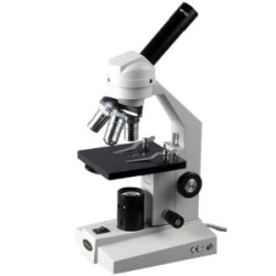


Features:
| Magnification | 25X - 1500x. |
| Observation | Binocular interchangeable with trinocular head for micro photography |
| Stand | Rectangular base heavy and stable with transformer and electrical fitted inside. |
| Stage | Fixed large size stage for inspection of large specimen with detachable graduated mechanical stage for X & Y movement of specimen. |
| Focusing | Coarse and fine focusing by conveniently placed separate knobs 1div = 0.002mm. |
| Illumination | Incident bright light through Epi-illuminator 12V-50W halogen lamp with iris diaphragm and filter slot.Continuously variable luminosity. |
| Nose Piece | Quadruple revolving nose piece with positive click stop. |
| Objectives | Achromatic - M5x, M10x, M20x, & M45x or M60x (S.L.) |
| Eyepieces | Huygenian 5x & wide filed 10x or 15x or 20x |
Colour Video coupler
- Digital signal processing camera with 1/3 image sensor. Automatic white balancing. Resolution 510 lines. Zoom lens for close micro photography.
Image Grabbing Card
Image grabber is designed to capture & display colour & grayscale video images. Trilinear MIP mapping, alpha blending, anti-aliasing, bi-linear filtering, gauraud shading makes the microphotography more realistic without losing scientific details of the object.
Software
Microcam Software is powerful for acquiring organising, storing, retrieving and editing any image.
The software provides nodules analysis, coating thickness measurement, depth or width measurement of decarburisation, phase percentage with display by coloured overlays, grain size no., porosity analysis, graphite flakes measurement, non - metallic inclusion rating, particle size and volume percentage with lot makes software complete. Generated reports can be saved or printed with images, histogram, data, overlays colours and various other related information.
MT-780 : Inverted Metallurgical Microscope
- Views: 456
-
 Just Machine Tools
 - Vendor Type: Manufacturer
- Product Code: JMT0000007
- Availability: In Stock
- ₹0