

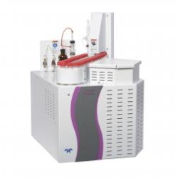


EEM® View -CMOS camera imaging system for Fluorescence Spectrophotometer-
Product Details :
EEM View is a completely new concept system in the world which delivers fluorescence, reflection spectra and these images simultaneously. To make it possible, AI technology is applied to analyze data with a special algorithm*1. This measurement is possible by installing the EEM View Accessory on the F-7000 / 7100 Fluorescent Spectrophotometer.
- The spectral analysis algorithm was developed in joint research by Professor Imari SATO and Associate Professor Yinqiang ZHENG of the National Institute of Informatics.
- "EEM" is a trademark of Hitachi High-Tech Science Corporation, registered in Japan and China.
What is EEM view?
New technology capable of capturing fluorescence and reflection images and spectra of a sample simultaneously.
- Measurement of spectrum data for samples (spectral and fluorescence properties).
- Captures images under white light or monochromatic light
(area: Φ20 mm, wavelength range: 380 to 700 nm)
- Displays separated fluorescence and reflection images obtained using an analysis algorithm that applies AI technology*1
- Displays spectra for each partition in an image*1
- (fluorescence spectrum and reflection spectrum)
- The spectral analysis algorithm was developed in joint research by Professor Imari SATO nd Associate Professor Yinqiang ZHENG of the National Institute of Informatics.
Overview
- Fluorescence Spectrophotometer equipped with CMOS camera imaging system -
Uniform illumination system using an integrating sphere
Captures fluorescence and reflection images and spectra of samples simultaneously!
- Diffusion of illumination using integrating sphere
- Highly uniform illumination of samples
- Dual detection using fluorescence spectroscope and CMOS camera
A spectrofluorometric microscope is an option that can be attached to the sample chamber of the existing Model F-7100 fluorescence spectrophotometer equipped with CMOS camera.
Simple mounting which supports many types of samples!
- Simple mounting means just placing the sample on top of the integrating sphere!
- Plate sample: Mount sample using the quartz window
- Powder sample: Fill the powder sample Petri dish with powder, and mount the sample. In addition, the powder cell included with the separate optional solid sample holder can be used.
- A fluorescence standard sample is included for alignment confirmation.
- The included reference white plate (100%) and blank plate (0%) are used to carry out calibration. These are used to calibrate the fluorescence intensity and the reflectance, and to correct the in-plane brightness distribution in the image.
EEM® View -CMOS camera imaging system for Fluorescence Spectrophotometer-
- Views: 184
-
 Aspire Inc
 - Vendor Type: Manufacturer, Supplier
- Product Code: KK00000055
- Availability: In Stock
- ₹0
Seller information